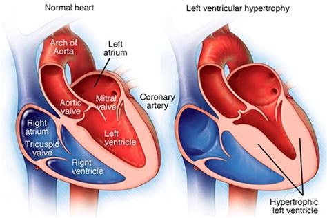
lv walls | normal Lv wall thickness lv walls Left ventricular hypertrophy is a thickening of the wall of the heart's main pumping chamber, called the left ventricle. This thickening may increase pressure within the heart. The condition can make it harder for the heart to pump blood. The most common cause is . Looking for information on the anime Lv2 kara Cheat datta Motoyuusha Kouho no Mattari Isekai Life (Chillin' in Another World with Level 2 Super Cheat Powers)? Find out more with MyAnimeList, the world's most active online anime and manga community and database.
0 · reasons for left ventricular hypertrophy
1 · normal Lv wall thickness
2 · myocardial wall
3 · lvh with repolarization abnormalities
4 · increased Lv wall thickness
5 · Lv wall thickness on echo
6 · Lv wall thickness normal values
7 · Lv wall motion abnormalities
MGM properties have endless things to do. Explore them all. Discover MGM. 3850 S Las Vegas Blvd. Las Vegas, NV 89109. Guest Services. Excalibur Hotel. MGM Rewards Mastercard. Receive Offers.
Segments of the left ventricle. Based on anatomical landmarks and autopsy studies (Edwards et al), the left ventricle is divided into three equal parts along . Recently, the consensus of the American Heart Association (AHA) 21 divided the LV into 4 walls: septal, anterior, lateral, and inferior; in turn, the .Segments of the left ventricle. Based on anatomical landmarks and autopsy studies (Edwards et al), the left ventricle is divided into three equal parts along the long axis of the ventricle. This creates three circular sections of the left ventricle named basal, mid-cavity, and apical. Recently, the consensus of the American Heart Association (AHA) 21 divided the LV into 4 walls: septal, anterior, lateral, and inferior; in turn, the 4 walls were divided into 17 segments: 6 basal, 6 mid, 4 apical, and 1 segment being the apex (Figure 2).
Left ventricular hypertrophy is a thickening of the wall of the heart's main pumping chamber, called the left ventricle. This thickening may increase pressure within the heart. The condition can make it harder for the heart to pump blood. The most common cause is .Assessment of LV function remains the most common reason for cardiac imaging because of its powerful ability to predict morbidity and mortality. Current routine methods of quantifying LV function (with LVEF) is not without limitations.Electronic calipers should be positioned on the interface between myocardial wall and cavity, and the interface between wall and pericardium. Perform at end-diastole (previously defined) perpendicular to the long axis of the LV, at or immediately below the level of .
Each echocardiogram includes an evaluation of the LV dimensions, wall thicknesses and function. Good measurements are essential and may have implications for therapy. The LV dimensions must be measured when the end-diastolic and end-systolic valves (MV and AoV) are closed in the parasternal long axis (PLAX) view. The first and most commonly used echocardiography method of LVM estimation is the linear method, which uses end-diastolic linear measurements of the interventricular septum (IVSd), LV inferolateral wall thickness, and LV internal diameter derived from 2D-guided M-mode or direct 2D echocardiography. This method utilizes the Devereux and Reichek .Each of the following echo parameters are discussed and updated in turn: left ventricular linear dimensions and LV mass; left ventricular volumes; left ventricular ejection fraction; left atrial size; right heart parameters; aortic dimensions; and tissue Doppler imaging.
reasons for left ventricular hypertrophy
Wall motion is assessed in each segment of the left ventricle (Figure 1; refer to Segments of the Left Ventricle). Regional wall motion abnormalities are defined as regional abnormalities in contractile function. Ischemic heart disease is the most common cause of .The volume-based measurement of left ventricular ejection fraction (LVEF) is fundamentally different from direct measurement of myocardial motion by tissue Doppler imaging and myocardial deformation, and the reliability and precision of these measurements are also different.Segments of the left ventricle. Based on anatomical landmarks and autopsy studies (Edwards et al), the left ventricle is divided into three equal parts along the long axis of the ventricle. This creates three circular sections of the left ventricle named basal, mid-cavity, and apical.
Recently, the consensus of the American Heart Association (AHA) 21 divided the LV into 4 walls: septal, anterior, lateral, and inferior; in turn, the 4 walls were divided into 17 segments: 6 basal, 6 mid, 4 apical, and 1 segment being the apex (Figure 2). Left ventricular hypertrophy is a thickening of the wall of the heart's main pumping chamber, called the left ventricle. This thickening may increase pressure within the heart. The condition can make it harder for the heart to pump blood. The most common cause is .
breitling quartz movement maintenance
Assessment of LV function remains the most common reason for cardiac imaging because of its powerful ability to predict morbidity and mortality. Current routine methods of quantifying LV function (with LVEF) is not without limitations.Electronic calipers should be positioned on the interface between myocardial wall and cavity, and the interface between wall and pericardium. Perform at end-diastole (previously defined) perpendicular to the long axis of the LV, at or immediately below the level of . Each echocardiogram includes an evaluation of the LV dimensions, wall thicknesses and function. Good measurements are essential and may have implications for therapy. The LV dimensions must be measured when the end-diastolic and end-systolic valves (MV and AoV) are closed in the parasternal long axis (PLAX) view. The first and most commonly used echocardiography method of LVM estimation is the linear method, which uses end-diastolic linear measurements of the interventricular septum (IVSd), LV inferolateral wall thickness, and LV internal diameter derived from 2D-guided M-mode or direct 2D echocardiography. This method utilizes the Devereux and Reichek .
Each of the following echo parameters are discussed and updated in turn: left ventricular linear dimensions and LV mass; left ventricular volumes; left ventricular ejection fraction; left atrial size; right heart parameters; aortic dimensions; and tissue Doppler imaging.Wall motion is assessed in each segment of the left ventricle (Figure 1; refer to Segments of the Left Ventricle). Regional wall motion abnormalities are defined as regional abnormalities in contractile function. Ischemic heart disease is the most common cause of .
normal Lv wall thickness
breitling premier panda dial

breitling pusher button replacement
Details. Low volume aggressive climbing shoe for bouldering and sport routes. Trax SAS rubber provides supreme friction on small holds and hooks. Neoflex neoprene covers Knuckle Box to retain stretch after flex. Hook-and-loop closure ensures easy on and off with simple adjustments. TPS+ maintains aggressive downturn overtime.
lv walls|normal Lv wall thickness


