lv end-diastolic dimensions should be measured at qrs | lv end diastolic measurements lv end-diastolic dimensions should be measured at qrs Aortic dimensions should be measured using 2D imaging from the PLAX window. Indices should be obtained using the inner-edge to inner-edge (IE-IE) methodology in end-diastole, defined . 4.7 129 ratings. Book 4 of 5: Villainess Level 99: I May Be the Hidden Boss but I'm Not the Demon Lord. See all formats and editions. After defeating the god of evil, Yumiella has finally surpassed the level cap of 99.
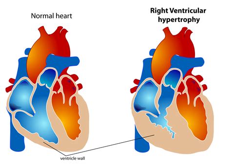
0 · right ventricular hypertrophy measurements
1 · lv heart size chart
2 · lv end diastolic measurements
3 · lv end diastolic
4 · left ventricular hypertrophy measurements
5 · left ventricular heart size chart
6 · left ventricular heart measurements
Material Matters: A Guide to Louis Vuitton Textiles. Whether you’re a seasoned collector or just starting to dabble in the world of designer brands, there’s a lot to learn (and appreciate) about the Louis Vuitton collection.
LV end-diastolic dimensions should always be measured the upstroke of the QRS. Partition values allow you separate the left atrium from the left ventricle. LV measurements taken from low parasternal windows overestimate true values. The diagnosis of LV hypertrophy is .Perform at end-diastole (previously defined) perpendicular to the long axis of the LV, at or immediately below the level of the mitral valve leaflet tips. LV mass = 0.8x (1.04x .Aortic dimensions should be measured using 2D imaging from the PLAX window. Indices should be obtained using the inner-edge to inner-edge (IE-IE) methodology in end-diastole, defined .Normal (reference) values for echocardiography, for all measurements, according to AHA, ACC and ESC, with calculators, reviews and e-book.
To measure end-diastolic diameter use the section of the MMode in which the ventricle is largest, shortly before the walls begin to move inward (onset of the QRS complex). For the end . The LV dimensions must be measured when the end-diastolic and end-systolic valves (MV and AoV) are closed in the parasternal long axis (PLAX) view. The measurement .LA volume should be measured at left ventricular end systole (largest LA size at the frame just before mitral valve opening) using the Simpson’s biplane MoD and then indexed to BSA. Since .QRS duration increased by 5.4 ms for every 100 g increase in LV mass, and by 4.6 ms for each 10 mm increase in LV end-diastolic diameter. The amplitude increased by 0.8 mm for every .
In patients with LBBB, LV length (r = 0.32, p = 0.03), mass (r = 0.39, p = 0.01), diameter (r = 0.34, p = 0.02), and LV end-diastolic volume (r = 0.32, p = 0.04) had positive . LV end-diastolic dimensions should always be measured the upstroke of the QRS. Partition values allow you separate the left atrium from the left ventricle. LV measurements taken from low parasternal windows overestimate true values. The diagnosis of LV hypertrophy is based on wall thickness. 2005.I will review the fundamentals of the correct techniques for accurate LV measurement, explain the timing of end diastole/systole in regards to linear measurements, discuss caliper location and outline 6 pitfalls to avoid when measuring the left ventricular wall and chambers.Perform at end-diastole (previously defined) perpendicular to the long axis of the LV, at or immediately below the level of the mitral valve leaflet tips. LV mass = 0.8x (1.04x [(IVS+LVID+PWT) 3 -LVID 3 ] + 0.6 grams
Aortic dimensions should be measured using 2D imaging from the PLAX window. Indices should be obtained using the inner-edge to inner-edge (IE-IE) methodology in end-diastole, defined as the onset of the QRS complex. All values should be indexed to height and not BSA.Normal (reference) values for echocardiography, for all measurements, according to AHA, ACC and ESC, with calculators, reviews and e-book.To measure end-diastolic diameter use the section of the MMode in which the ventricle is largest, shortly before the walls begin to move inward (onset of the QRS complex). For the end-systolic dimension, pick the region in which the ventricular cavity is smallest.
The LV dimensions must be measured when the end-diastolic and end-systolic valves (MV and AoV) are closed in the parasternal long axis (PLAX) view. The measurement is performed in the basal portion of the LV by the chordae. Left ventricular dimensions. Left ventricular geometry and mass. References.
right ventricular hypertrophy measurements
LA volume should be measured at left ventricular end systole (largest LA size at the frame just before mitral valve opening) using the Simpson’s biplane MoD and then indexed to BSA. Since apical views that are optimised for the LV will foreshorten the LA, dedicated apical 4-chamber and 2-chamber images should be acquired to maximise LA .QRS duration increased by 5.4 ms for every 100 g increase in LV mass, and by 4.6 ms for each 10 mm increase in LV end-diastolic diameter. The amplitude increased by 0.8 mm for every 100 g increase in LV mass. In patients with LBBB, LV length (r = 0.32, p = 0.03), mass (r = 0.39, p = 0.01), diameter (r = 0.34, p = 0.02), and LV end-diastolic volume (r = 0.32, p = 0.04) had positive correlations with QRSd.
LV end-diastolic dimensions should always be measured the upstroke of the QRS. Partition values allow you separate the left atrium from the left ventricle. LV measurements taken from low parasternal windows overestimate true values. The diagnosis of LV hypertrophy is based on wall thickness. 2005.I will review the fundamentals of the correct techniques for accurate LV measurement, explain the timing of end diastole/systole in regards to linear measurements, discuss caliper location and outline 6 pitfalls to avoid when measuring the left ventricular wall and chambers.Perform at end-diastole (previously defined) perpendicular to the long axis of the LV, at or immediately below the level of the mitral valve leaflet tips. LV mass = 0.8x (1.04x [(IVS+LVID+PWT) 3 -LVID 3 ] + 0.6 gramsAortic dimensions should be measured using 2D imaging from the PLAX window. Indices should be obtained using the inner-edge to inner-edge (IE-IE) methodology in end-diastole, defined as the onset of the QRS complex. All values should be indexed to height and not BSA.
Normal (reference) values for echocardiography, for all measurements, according to AHA, ACC and ESC, with calculators, reviews and e-book.To measure end-diastolic diameter use the section of the MMode in which the ventricle is largest, shortly before the walls begin to move inward (onset of the QRS complex). For the end-systolic dimension, pick the region in which the ventricular cavity is smallest. The LV dimensions must be measured when the end-diastolic and end-systolic valves (MV and AoV) are closed in the parasternal long axis (PLAX) view. The measurement is performed in the basal portion of the LV by the chordae. Left ventricular dimensions. Left ventricular geometry and mass. References.LA volume should be measured at left ventricular end systole (largest LA size at the frame just before mitral valve opening) using the Simpson’s biplane MoD and then indexed to BSA. Since apical views that are optimised for the LV will foreshorten the LA, dedicated apical 4-chamber and 2-chamber images should be acquired to maximise LA .
QRS duration increased by 5.4 ms for every 100 g increase in LV mass, and by 4.6 ms for each 10 mm increase in LV end-diastolic diameter. The amplitude increased by 0.8 mm for every 100 g increase in LV mass.
lv heart size chart

lv end diastolic measurements
lv end diastolic
June 05, 2020. Drug Prevention. DEA's revised and updated drug fact sheet about MDMA, also known as ecstasy - what it is, what is its origin, what common street names for these drugs, what does it look like, how it is abused, what their effect is on the mind and bodies of users including signs of overdose, and its legal status. Download.
lv end-diastolic dimensions should be measured at qrs|lv end diastolic measurements


