lv summit anatomy | lmca anatomy diagram lv summit anatomy We provide a systematic atlas and nomenclature of LV summit veins . This pre-owned OMEGA Seamaster 300M 2223.80.00 watch is in very good condition. During our quality control check, any necessary adjustments are made to ensure the .
0 · right ventricular summit arrhythmia map
1 · lvs diagram
2 · lvs anatomy
3 · lmca anatomy diagram
4 · left ventricular summit arrhythmia diagram
5 · anatomy of the left ventricular
6 · anatomy of left ventricular summit
The Cartier Tank Mini LC measures 24mm x 16.5mm, is fitted with a quartz movement, and is water resistant up to 30 meters. Priced at $7,000, launching in .
right ventricular summit arrhythmia map
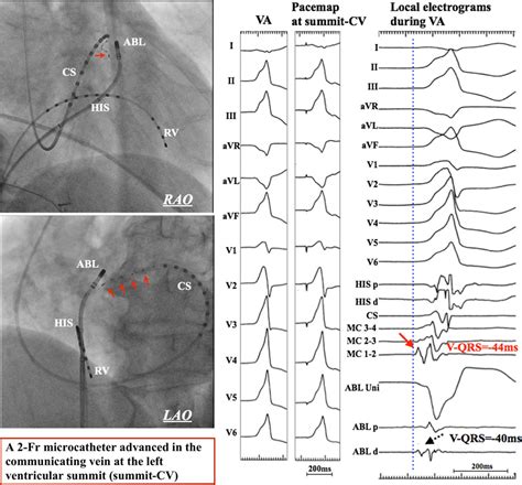
The left ventricular summit (LVS) is a triangular area located at the most superior portion of the left epicardial ventricular region, surrounded by the two branches of the left .We provide a systematic atlas and nomenclature of LV summit veins .
This article presents a review of the anatomy of the LVS and relevant .
men cartier ring
Results: Angiography identified the following LVS veins: (1) LV annular . In this article, we review the anatomy of the LVS and our approach to mapping and ablating arrhythmias originating from this region. The summit of the left ventricle (LV) is the most superior portion of the epicardial LV bounded by an arc from the left anterior descending coronary artery, superior to the first . Left ventricular summit (LVS) arrhythmias are challenging as catheter ablation targets. This review summarizes LVS anatomy, mapping techniques, and endocardial and .
We provide a systematic atlas and nomenclature of LV summit veins related to arrhythymogenic substrates. vCTA can be useful for noninvasive evaluation of LV summit veins prior to ethanol . This article presents a review of the anatomy of the LVS and relevant regions and discusses novel mapping and ablation techniques for eliminating LVS ventricular arrhythmias.
The LV summit is the most superior portion of the LV (star, B) and an important anatomic landmark as it is the region on the epicardial surface, where the left main coronary .The left ventricular (LV) summit (LVS) is the most septal and superior aspect of the left ventricular outflow tract (LVOT), bound superiorly and anteriorly by the left main coronary artery (LMCA) bifurcation and laterally by the great cardiac vein .
LV VAs.2 The complex relationships between the left ventricular summit (LVS) and surrounding structures under-score the importance of understanding the anatomy of this region and the .Results: Angiography identified the following LVS veins: (1) LV annular branch of the great cardiac vein (GCV) (19/53); (2) septal (rightward) branches of the anterior ventricular vein (AIV) . The left ventricular summit (LVS) is a triangular area located at the most superior portion of the left epicardial ventricular region, surrounded by the two branches of the left coronary artery: the left anterior interventricular artery and the left circumflex artery. In this article, we review the anatomy of the LVS and our approach to mapping and ablating arrhythmias originating from this region.
The summit of the left ventricle (LV) is the most superior portion of the epicardial LV bounded by an arc from the left anterior descending coronary artery, superior to the first septal perforating branch to the left circumflex coronary artery. Left ventricular summit (LVS) arrhythmias are challenging as catheter ablation targets. This review summarizes LVS anatomy, mapping techniques, and endocardial and epicardial ablation techniques. Advanced ablation strategies are increasingly being developed to access intramural substrate and improve procedural success.
We provide a systematic atlas and nomenclature of LV summit veins related to arrhythymogenic substrates. vCTA can be useful for noninvasive evaluation of LV summit veins prior to ethanol ablation.
This article presents a review of the anatomy of the LVS and relevant regions and discusses novel mapping and ablation techniques for eliminating LVS ventricular arrhythmias.
The LV summit is the most superior portion of the LV (star, B) and an important anatomic landmark as it is the region on the epicardial surface, where the left main coronary artery (LMCA) bifurcates and is recognized as the commonest source of idiopathic epicardial ventricular arrhythmias (VAs). 35 Anatomic landmarks defining the LV summit are .The left ventricular (LV) summit (LVS) is the most septal and superior aspect of the left ventricular outflow tract (LVOT), bound superiorly and anteriorly by the left main coronary artery (LMCA) bifurcation and laterally by the great cardiac vein (GCV). 1–3 Arrhythmias arising in LVS pose a challenge to ablation because catheter manipulation .LV VAs.2 The complex relationships between the left ventricular summit (LVS) and surrounding structures under-score the importance of understanding the anatomy of this region and the value of imaging techniques for detailed mapping and safe ablation. In this article, we review the anatomy of the LVS and our approach to mapping andResults: Angiography identified the following LVS veins: (1) LV annular branch of the great cardiac vein (GCV) (19/53); (2) septal (rightward) branches of the anterior ventricular vein (AIV) (53/53); and (3) diagonal branches of the AIV (51/53).

The left ventricular summit (LVS) is a triangular area located at the most superior portion of the left epicardial ventricular region, surrounded by the two branches of the left coronary artery: the left anterior interventricular artery and the left circumflex artery. In this article, we review the anatomy of the LVS and our approach to mapping and ablating arrhythmias originating from this region. The summit of the left ventricle (LV) is the most superior portion of the epicardial LV bounded by an arc from the left anterior descending coronary artery, superior to the first septal perforating branch to the left circumflex coronary artery.
Left ventricular summit (LVS) arrhythmias are challenging as catheter ablation targets. This review summarizes LVS anatomy, mapping techniques, and endocardial and epicardial ablation techniques. Advanced ablation strategies are increasingly being developed to access intramural substrate and improve procedural success.We provide a systematic atlas and nomenclature of LV summit veins related to arrhythymogenic substrates. vCTA can be useful for noninvasive evaluation of LV summit veins prior to ethanol ablation.
This article presents a review of the anatomy of the LVS and relevant regions and discusses novel mapping and ablation techniques for eliminating LVS ventricular arrhythmias. The LV summit is the most superior portion of the LV (star, B) and an important anatomic landmark as it is the region on the epicardial surface, where the left main coronary artery (LMCA) bifurcates and is recognized as the commonest source of idiopathic epicardial ventricular arrhythmias (VAs). 35 Anatomic landmarks defining the LV summit are .
The left ventricular (LV) summit (LVS) is the most septal and superior aspect of the left ventricular outflow tract (LVOT), bound superiorly and anteriorly by the left main coronary artery (LMCA) bifurcation and laterally by the great cardiac vein (GCV). 1–3 Arrhythmias arising in LVS pose a challenge to ablation because catheter manipulation .LV VAs.2 The complex relationships between the left ventricular summit (LVS) and surrounding structures under-score the importance of understanding the anatomy of this region and the value of imaging techniques for detailed mapping and safe ablation. In this article, we review the anatomy of the LVS and our approach to mapping and

cartier bracelet white gold
Gladfield Ale Malt 25kg. In stock. Add to Cart. $ 85.00. Gladfield Ale malt is made from Autumn grown 2-row barley varieties. The malt is fully modified through a traditional long .
lv summit anatomy|lmca anatomy diagram


